Biophysics explains how immune cells kill bacteria

A new data analysis technique, moving subtrajectory analysis defines the dynamics and kinetics of key molecules in the immune response to an infection. These biophysical descriptions are expected to clarify the TCR microcluster, an essential assembly for a T cell to initiate its attack on a pathogen.

CD3?-EGFP (green), Qdot 655-labeled CD3? (red), and Qdot 585-labeled CD45 (blue) in living Jurkat cells.

A new data analysis technique, moving subtrajectory analysis, designed by researchers at Tokyo Institute of Technology, defines the dynamics and kinetics of key molecules in the immune response to an infection. These biophysical descriptions are expected to clarify the TCR microcluster, an essential assembly for a T cell to initiate its attack on a pathogen.
To kill a pathogen invading the human body, T cells, or lymphocytes, bind to it through T cell receptors (TCR). One of the first events this binding initiates is the formation of a microcluster that includes tens or hundreds of TCR molecules. These microclusters are deemed essential to initiate and sustain the immune signal. A new analysis technique by scientists at School of Life Sciences at Tokyo Institute of Technology provides a quantitative description of the molecules that form these microclusters. The study can be read in Scientific Reports.
"Imaging technologies have visualized the generation and dynamics of microclusters, but there is no quantitative data. We developed 'moving subtrajectory (MST) analysis' using single-molecule tracking to quantitatively study the dynamics and kinetics of CD3 and CD45 around the microcluster," explains Prof. Makio Tokunaga, whose lab designed the new method.
TCR function by forming a complex (TCR/CD3) with CD3. CD45, on the other hand, is not part of the complex, but is believed to regulate the formation of the cluster.
Single molecule imaging was used to trace the movement of CD3 and CD45 around the microclusters. Yuma Ito, an Assistant Professor in the lab, shows that MST analysis is superior to standard analysis methods by revealing details on the temporal and spatial variation of the movement.
"Standard methods analyze the mean square displacement of the whole trajectory. MST divides the trajectory into subtrajectories and calculates the mean square displacement of each subtrajectory. Using MST, we could analyze movement inside, outside and at the boundary of the TCR microclusters," he explains.
The result yields much more data that can be used to parse information about CD3 and CD45 behavior during the creation of TCR microclusters. The researchers found that the dynamics of the two molecules depended on their location relative to the microclusters and could be used to determine their interactions with the microclusters. 
The kinetics showed CD3 and CD45 could take either a fast or slow mobility state. Inside the microcluster, the slow mobility state was dominant, reflecting stronger interactions between the two molecules and the microcluster. Outside and at the boundary, however, the fast mobility state was dominant. Yet a small fraction of molecules behaved according to the slow mobility state, suggesting that TCR nanoclusters exist at the outside or boundary of the microcluster.
"The appearance of nanoclusters gives us a new understanding of the dynamics of microclusters. The dynamics and kinetics at the single molecule level is very important for defining the molecular mechanisms of biological functions. Along with the new development of biopharmaceuticals related to immune control, elucidation of the mechanism of T cell activation is becoming more important in clinical application. Detailed quantitative data will assist in manufacturing biopharmaceuticals with stronger effects on the immune system," says Tokugawa.
While the paper demonstrates the application of MST analysis to TCR microclusters, the quantitative tool is applicable to any cellular complex that depends on spatiotemporal dynamics, let alone those of the immune system.

Eating habits affect skin's protection against sun

Sunbathers may want to avoid midnight snacks before catching some rays, new research recommends. A study in mice shows that eating at abnormal times disrupts the biological clock of the skin, including the daytime potency of an enzyme that protects against the sun's harmful ultraviolet radiation.


                                                 This is a skin cancer cell.


Sunbathers may want to avoid midnight snacks before catching some rays.
A study in mice from the O'Donnell Brain Institute and UC Irvine shows that eating at abnormal times disrupts the biological clock of the skin, including the daytime potency of an enzyme that protects against the sun's harmful ultraviolet radiation.
Although further research is needed, the finding indicates that people who eat late at night may be more vulnerable to sunburn and longer-term effects such as skin aging and skin cancer, said Dr. Joseph S. Takahashi, Chairman of Neuroscience at UT Southwestern Medical Center's Peter O'Donnell Jr. Brain Institute.
"This finding is surprising. I did not think the skin was paying attention to when we are eating," said Dr. Takahashi, also an Investigator with the Howard Hughes Medical Institute.
The study showed that mice given food only during the day -- an abnormal eating time for the otherwise nocturnal animals -- sustained more skin damage when exposed to ultraviolet B (UVB) light during the day than during the night. This outcome occurred, at least in part, because an enzyme that repairs UV-damaged skin -- xeroderma pigmentosum group A (XPA) -- shifted its daily cycle to be less active in the day.
Mice that fed only during their usual evening times did not show altered XPA cycles and were less susceptible to daytime UV rays.
"It is likely that if you have a normal eating schedule, then you will be better protected from UV during the daytime," said Dr. Takahashi, holder of the Loyd B. Sands Distinguished Chair in Neuroscience. "If you have an abnormal eating schedule, that could cause a harmful shift in your skin clock, like it did in the mouse."
Previous studies have demonstrated strong roles for the body's circadian rhythms in skin biology. However, little had been understood about what controls the skin's daily clock.
The latest research published in Cell Reports documents the vital role of feeding times, a factor that scientists focused on because it had already been known to affect the daily cycles of metabolic organs such as the liver.
The study found that besides disrupting XPA cycles, changing eating schedules could affect the expression of about 10 percent of the skin's genes.
However, more research is needed to better understand the links between eating patterns and UV damage in people, particularly how XPA cycles are affected, said Dr. Bogi Andersen of University of California, Irvine, who led the collaborative study with Dr. Takahashi.
"It's hard to translate these findings to humans at this point," said Dr. Andersen, Professor of Biological Chemistry. "But it's fascinating to me that the skin would be sensitive to the timing of food intake."
Dr. Takahashi, noted for his landmark discovery of the Clock gene regulating circadian rhythms, is researching other ways in which eating schedules affect the biological clock. A study earlier this year reinforced the idea that the time of day food is eaten is more critical to weight loss than the amount of calories ingested. He is now conducting long-term research measuring how feeding affects aging and longevity.
The UV study was supported by the Irving Weinstein Foundation, the National Institutes of Health, the China Scholarship Council, and the National Science Foundation Graduate Research Fellowship.

Problems with DNA replication can cause epigenetic changes that may be inherited for several generations

Scientists reveal that a fault in the process that copies DNA during cell division can cause epigenetic changes that may be inherited for up-to five generations. They also identified the cause of these epigenetic changes, which is related to the loss of a molecular mechanism in charge of silencing genes. Their results will change the way we think about the impact of replication stress in cancer and during embryonic development, as well as its inter-generational inheritance.


Scientists reveal that a fault in the process that copies DNA during cell division can cause epigenetic changes that may be inherited for up-to five generations. They also identified the cause of these epigenetic changes, which is related to the loss of a molecular mechanism in charge of silencing genes. Their results, which will be published in Science Advances on 16 August, will change the way we think about the impact of replication stress in cancer and during embryonic development, as well as its inter-generational inheritance.
Cell division is key for renewing the cells in our tissues and organs. There are two particular processes in which cell division is crucial: embryonic development and tumorigenesis. A fault in the process that copies DNA during cell division can cause genetic changes, so impaired DNA replication is a well-known cancer hallmark and a driver of genomic instability.
Now, scientists at the Centre for Genomic Regulation (CRG) in collaboration with the Josep Carreras Leukaemia Research Institute (IJC) and The Institute for Health Science Research Germans Trias i Pujol (IGTP) have discovered that impaired DNA replication can also cause large epigenetic changes. Their study, which was performed in worms (the model organism Caenorhabditis elegans), suggests that these genome-wide epigenetic alterations establish new gene expression states that may be inherited for up-to five generations. 
This is a striking example of trans-generational inheritance of epigenetic changes meaning that two individuals may differ in gene expression only because of the stress experienced by their ancestors.
The researchers, led by ICREA research professor and group leader at the CRG Ben Lehner, also identified the mechanism causing these epigenetic changes. "For the correct function of cells and ultimately the health of the organism, it is important to keep certain genes active and others silenced. Inside cells, there are DNA-protein complexes called heterochromatin that prevent genes from becoming activated when they should not be. Initially, 
we noticed that a gene artificially inserted into the worm genome and normally silenced by heterochromatin was activated in animals that carried mutations in proteins involved in the copying of DNA," explains Tanya Vavouri CRG alumna currently group leader at IJC and IGTP and coauthor of this study. "We found that this was caused by loss of heterochromatin and that other genes also silenced by heterochromatin were activated too. Unexpectedly, the gene was inappropriately activated for five generations in animals that did not carry the mutation in DNA replication but had ancestors that did," she adds.
"Our results show that impaired DNA replication not only causes genetic alterations but also genome-wide epigenetic changes that can be stably inherited," says Ben Lehner, senior author of the paper. 
An important question in epigenetics is the extent to which epigenetic states are transmitted between generations. Lehner and collaborators are addressing this and other questions from many different angles. 
They previously reported that some temperature-induced gene expression changes can also be inherited between generations. "We hope that our work will change the way people think about the impact of replication stress during tumorigenesis and embryonic development as well as about inter-generational inheritance," he concludes.

Tasmanian devils begin to resist infectious cancer

Genetic tweaks are helping the Australian species ward off the killer disease

A contagious facial tumor has killed up to 95 percent of Tasmanian devils in some locations. But genetic variants in a small number of devils may let them avoid the disease. This discovery could be good news for the species.

A contagious cancer has been killing off Tasmanian devils in large numbers. But some have avoided the disease. The key to their salvation, a new study shows, has been their genes.
Tasmanian devils are small, aggressive marsupials on the Australian island of Tasmania. In 1996, researchers noticed some devils had tumors on their faces. Those cancers can stop an animal from eating and breathing, causing death. Unlike most human cancers, this one can spread from animal to animal. The infectious disease was first traced to a female devil. It spread its cancer to other devils through biting. (These animals frequently bite each other during mating.) The disease has now wiped out about 80 percent of the fierce animals. In some places, it has killed up to 95 percent of them.
Usually cells from different animals can’t grow in each other’s’ bodies. If you kiss your mom, her cells don’t start growing on your lips. The immune system, which helps fight off germs, also kills cells from other individuals. That is why people who get organ transplants have to take drugs to stop their bodies from rejecting those “foreign” organs. But devil facial tumors can hide from the immune system.
Tasmanian devil cancer
The cancer causes tumors on the face of an infected Tasmanian devil. Those tumors can interfere with eating and breathing.
This was one reason scientists worried about the devils. “What we reluctantly felt was that this was the end for the Tasmanian devil,” says Jim Kaufman. There were worries it could go extinct “because they really didn’t have a defense.” Kaufman is a scientist who studies how the immune system evolved. He works at the University of Cambridge in England.
But researchers have just turned up good news. Many devils have versions of genes that may protect them from the cancer. Researchers reported this August 30 in Nature Communications. Concludes Kaufman, this “is really the most hopeful thing I’ve heard in a long time.”

In the genes

Menna Jones is a conservation biologist at the University of Tasmania in Hobart. As the cancer spread across Tasmania, she and her coworkers collected DNA from devils in three places. They did this both before and after the disease arrived at the locations.
Jones then teamed up with Andrew Storfer. He is an evolutionary geneticist from Washington State University in Pullman. His team examined the devil’s genetic instruction book, or genome. These researchers wanted to see if there was something special about devils who remained healthy after the infectious disease arrived. That might explain why they survived while the rest did not.
Some scientists had suspected the survivors just hadn’t caught the cancer yet, Kaufman notes. It was thought they had been too young to breed and get bitten. The new study, though, indicates that differences in their DNA may have protected some devils from the cancer.
Storfer and his team found more than 90,000 DNA spots where a small number of devils have a different base — an information-carrying component of DNA — than in most devils. These genetic spelling differences are known as single nucleotide polymorphisms (PAH-lee-MORF-isms), or SNPs, pronounced “snips.” The team looked for SNPs that had been rare before the tumor swept through a population but then became common after the disease arrived. Such a pattern could indicate that natural selection was working. This evolutionary process meant devils with the right SNP variants would avoid the deadly infection.
Two parts of the genome in all three of the devil populations contained SNPs that fit such a profile. Because the two regions changed in all three populations, the change probably didn’t happen by chance, the researchers say. These variants aren’t new. They were already there. But very few devils had them before the tumor came. What changed? Only devils with those SNPs survived long enough to breed and have babies. And their offspring all inherited the protective SNPs.
Those two genome regions contain a total of seven genes. Genes are the specific instructions in the big genome instruction book. They tell cells how to build particular proteins. Proteins do most of the work of building cells, digesting food and all the other things a body must do. Some genes that fight the facial tumor have, in other mammals, been shown to combat cancer or control the immune system. The researchers, however, aren’t sure which of the genes were most protective for the devils, let alone how they might work.
Tasmania map
A facial tumor that kills Tasmanian devils was first discovered in 1996. Its female victim lived in the northeastern tip of the island of Tasmania. Since then, the disease has swept across the Australian island (red line shows how far the disease had spread by 2000, 2005, 2010 and 2015). In a new study, researchers collected DNA from devils at three sites (large red circles) before and after the infectious cancer reached them. (Additional sampling sites are shown in gray. Dates in parentheses indicate when disease reached the main sample sites.)
One day, scientists might use the new SNP data to better predict which devils will be at risk of the deadly face cancer. Breeding programs set up to save the devils might also use these data. Such programs might mate animals that carry the survival variants to ones that don’t. That would up the chance that the next generation would carry the guardian SNPs.
But there is a new complication. Last year, a second devil facial cancer emerged. Also infectious, it looks very much like the first tumor. In this case, it started in a male. Katherine Belov is a comparative genomicist at the University of Sydney in Australia. Researchers don’t know whether the SNPs that allow devils to resist the first infectious cancer will also work against the second, he notes. That’s why conservation biologists should not breed the resistance genes into all Tasmanian devils, she warns.
Limiting which devils breed with which others could reduce the diversity of genes in this species. Genetic diversity is high when there are many versions of genes present throughout members of a species. And devils need all the diversity they can get to cope with other diseases and other unknown challenges in the future, Belov says. Restricting breeding to animals that have these anti-tumor SNPs could limit the species’ ability to overcome other problems in the future.

Cancers that spread

Several cancer can be spread by infectious microbes, such as viruses. Feline leukemia is a well-known example. But what afflicts the devils is different. It’s not a germ that infects them but a sickening cell — a cancer cell — from a member of their own species.
Scientists used to think that such contagious cancer cells were rare. For a long time, only one type was known — in dogs. For 11,000 years, some of them had been spreading such an infectious dog cancer through mating. Then the first contagious devil cancer emerged 20 years ago. Scientists reported the second one last year.
But it was far from the only such new infectious cancer. Also in 2015, researchers found such a cancer — in this case, a leukemia — infecting soft shell clams. This past June, researchers reported finding more contagious shellfish cancers. They infected mussels, cockles and golden shell clams. Golden shell clams (Polititapes aureus) caught their cancer from pullet carpet shell clams (Venerupis corrugate), which no longer carry the disease. That was the first report of such contagious cancer cells infecting a new species.
With two such infectious cancers in Tasmanian devils, some researchers now wonder if their species is especially prone to contagious cancers. No one can yet answer that.
But in more good news for the devils, scientists have reared many healthy animals in captivity. Last month, 33 of them were released into the wild. Those devils had been given vaccines to help them ward off the facial tumor. Scientists hope they will breed with wild devils remaining to increase their genetic diversity.
The researchers have outfitted the newly released devils with collars with tracking “tags.” This will help them map the animals as they move around. The collars also are reflective. That may help to save these animals from the fate of devils previously released: Many were hit and killed by cars.
breed     (noun) Animals within the same species that are so genetically similar that they produce reliable and characteristic traits. German shepherds and dachshunds, for instance, are examples of dog breeds. (verb) To produce offspring through reproduction.
cancer     Any of more than 100 different diseases, each characterized by the rapid, uncontrolled growth of abnormal cells. The development and growth of cancers, also known as malignancies, can lead to tumors, pain and death.
cell     The smallest structural and functional unit of an organism. Typically too small to see with the naked eye, it consists of watery fluid surrounded by a membrane or wall. Animals are made of anywhere from thousands to trillions of cells, depending on their size. Some organisms, such as yeasts, molds, bacteria and some algae, are composed of only one cell.
cockles     A type of clam that with a somewhat ribbed, triangular shell. These mollusks have a strong, muscular foot that allows them to effectively hop along the ocean floor when they need to move. They tend to burrow just below the mud or sand in tidal beach areas and to depths of 15 meters (50 feet).
colleague     Someone who works with another; a co-worker or team member.
component     An item that is part of something else, such as pieces that go on an electronic circuit board.
conservation     The act of preserving or protecting something. The focus of this work can range from art objects to endangered species and other aspects of the natural environment.
conservation biologist     A scientist who investigates strategies for helping preserve ecosystems and especially species that are in danger of extinction.
contagious     Likely to infect or spread to others through direct or indirect contact; infectious.
defense     (in biology) A natural protective action taken or chemical response that occurs when a species confront predators or agents that might harm it. (adj. defensive)
diversity     (in biology) A range of different life forms.
evolutionary     An adjective that refers to changes that occur within a species over time as it adapts to its environment. Such evolutionary changes usually reflect genetic variation and natural selection, which leave a new type of organism better suited for its environment than its ancestors. The newer type is not necessarily more “advanced,” just better adapted to the conditions in which it developed.
gene     (adj. genetic) A segment of DNA that codes, or holds instructions, for producing a protein. Offspring inherit genes from their parents. Genes influence how an organism looks and behaves.
genetic     Having to do with chromosomes, DNA and the genes contained within DNA. The field of science dealing with these biological instructions is known as genetics. People who work in this field are geneticists.
genetic diversity     The range of genes types — and traits — within a population.
genome     The complete set of genes or genetic material in a cell or an organism. The study of this genetic inheritance housed within cells is known as genomics.
germ     Any one-celled microorganism, such as a bacterium, fungal species or virus particle. Some germs cause disease. Others can promote the health of higher-order organisms, including birds and mammals. The health effects of most germs, however, remain unknown.
immune system     The collection of cells and their responses that help the body fight off infections and deal with foreign substances that may provoke allergies.
infectious      An adjective that describes a type of germ that can be transmitted to people, animals or other living things.
leukemia     A type of cancer in which the bone marrow makes high numbers of immature or abnormal white blood cells. This can lead to anemia, a shortage of red blood cells.
mammal     A warm-blooded animal distinguished by the possession of hair or fur, the secretion of milk by females for feeding the young, and (typically) the bearing of live young.
marsupials     Mammals that carry their young for a period after birth in external pouches where the developing babies will have access to their mother’s nipples — and milk. Most of these species evolved in Australian and have especially long hind-legs. Examples of marsupials include kargaroos, opossums and koalas.
natural selection     This is guiding concept underlying evolution, or natural adaptation. It holds that natural mutations within a population of organisms will create some new forms that are better adapted to their environment. That adaptation makes them more likely to survive and reproduce. Over time, these survivors may come to dominate the original population. If their adaptive changes are significant enough, those survivors may also constitute a new species.
organ     (in biology) Various parts of an organism that perform one or more particular functions. For instance, an ovary is an organ that makes eggs, the brain is an organ that interprets nerve signals and a plant’s roots are organs that take in nutrients and moisture.
population     (in biology) A group of individuals from the same species that lives in the same area.
proteins     Compounds made from one or more long chains of amino acids. Proteins are an essential part of all living organisms. They form the basis of living cells, muscle and tissues; they also do the work inside of cells. The hemoglobin in blood and the antibodies that attempt to fight infections are among the better-known, stand-alone proteins. Medicines frequently work by latching onto proteins.
resistance     (as in drug resistance) The reduction in the effectiveness of a drug to cure a disease, usually a microbial infection. (as in disease resistance) The ability of an organism to fight off disease. (as in exercise) A type of rather sedentary exercise that relies on the contraction of muscles to build strength in localized tissues.
single nucleotide polymorphism (or SNP)     A SNP (pronounced “snip”) is DNA in which one of its original nucleotides has been naturally substituted for another. This variation may alter the function of DNA. SNPs are inherited. Each person carries millions of SNPs, making them unique from other people.
species     A group of similar organisms capable of producing offspring that can survive and reproduce.
tag      (in biology) To attach some rugged band or package of instruments onto an animal. Sometimes the tag is used to give each individual a unique identification number. Once attached to the leg, ear or other part of the body of a critter, it can effectively become the animal’s “name.” In some instances, a tag can collect information from the environment around the animal as well. This helps scientists understand both the environment and the animal’s role within it.
tumor     A mass of cells characterized by atypical and often uncontrolled growth. Benign tumors will not spread; they just grow and cause problems if they press against or tighten around healthy tissue. Malignant tumors will ultimately shed cells that can seed the body with new tumors. Malignant tumors are also known as cancers.
vaccine     A biological mixture that resembles a disease-causing agent. It is given to help the body create immunity to a particular disease. The injections used to administer most vaccines are known as vaccinations.
variant     A version of something that may come in different forms. (in biology) Members of a species that possess some feature (size, coloration or lifespan, for example) that make them distinct. (in genetics) A gene having a slight mutation that may have left its host species somewhat better adapted for its environment.

A new tool could one day improve Lyme disease diagnosis

‘Fingerprint’ test distinguishes between two easily confused tick-borne illnesses


A new testing method can distinguish between early Lyme disease and a similar tick-borne illness, researchers report. The approach may one day lead to a reliable diagnostic test for Lyme, an illness that can be challenging to identify.

Using patient blood serum samples, the test accurately discerned early Lyme disease from the similar southern tick‒associated rash illness, or STARI, up to 98 times out of 100. When the comparison also included samples from healthy people, the method accurately identified early Lyme disease up to 85 times out of 100, beating a commonly used Lyme test’s rate of 44 of 100, researchers report online August 16 in Science Translational Medicine. The test relies on clues found in the rise and fall of the abundance of molecules that play a role in the body’s immune response.
“From a diagnostic perspective, this may be very helpful, eventually,” says Mark Soloski, an immunologist at Johns Hopkins Medicine who was not involved with the study. “That’s a really big deal,” he says, especially in areas such as the mid-Atlantic where Lyme and STARI overlap.

In the United States, Lyme disease is primarily caused by an infection with the bacteria Borrelia burgdorferi, which is spread by the bite of a black-legged tick. An estimated 300,000 cases of Lyme occur nationally each year. Patients usually develop a rash and fever, chills, fatigue and aches. Black-legged ticks live in the northeastern, mid-Atlantic and north-central United States, and the western black-legged tick resides along the Pacific coast.

An accurate diagnosis can be difficult early in the disease, says immunologist Paul Arnaboldi of New York Medical College in Valhalla, who was not involved in the study. Lyme disease is diagnosed based on the rash, symptoms and tick exposure. But other illnesses have similar symptoms, and the rash can be missed. 

A test for antibodies to the Lyme pathogen can aid diagnosis, but it works only after a patient has developed an immune response to the disease.

STARI, spread by the lone star tick, can begin with a rash and similar, though typically milder, symptoms. The pathogen responsible for STARI is still unknown, though B. burgdorferi has been ruled out. So far STARI has not been tied to arthritis or other chronic symptoms linked to Lyme, though the lone star tick has been connected to a serious allergy to red meat (SN: 8/19/17, p. 16). Parts of both ticks’ ranges overlap, adding to diagnosis difficulties.

John Belisle, a microbiologist at Colorado State University in Fort Collins, and his colleagues had previously shown that a testing method based on small molecules related to metabolism could distinguish between early Lyme disease and healthy serum samples. “Think of it as a fingerprint,” he says. 

The method takes note of differences in the abundancy of metabolites, such as sugars, lipids and amino acids, involved in inflammation.
In the new work, Belisle and colleagues measured differences in the levels of metabolites in serum samples from Lyme and STARI patients. 

The researchers then developed a “fingerprint” based on 261 small molecules to differentiate between the two illnesses. To determine the accuracy, they tested another set of samples from patients with Lyme and STARI as well as those from healthy people. “We were able to distinguish all three groups,” says Belisle.

As a diagnostic test, “I think the approach has promise,” says Arnaboldi. But more work will be necessary to see if the method can sort out early Lyme disease, STARI and other tick-borne diseases in patients with unknown illnesses.

Having information about the metabolites abundant in STARI may also help researchers learn more about this disease, says Soloski. “This is going to spur lots of future studies.”

Bacteria genes offer new strategy for sterilizing mosquitoes

Genetic engineering could deplete populations of disease-carrying insects


A pair of bacterial genes may enable genetic engineering strategies for curbing populations of virus-transmitting mosquitoes.

Bacteria that make the insects effectively sterile have been used to reduce mosquito populations. Now, two research teams have identified genes in those bacteria that may be responsible for the sterility, the groups report online February 27 in Nature and Nature Microbiology.

“I think it’s a great advance,” says Scott O’Neill, a biologist with the Institute of Vector-Borne Disease at Monash University in Melbourne, Australia. People have been trying for years to understand how the bacteria manipulate insects, he says.

Wolbachia bacteria “sterilize” male mosquitoes through a mechanism called cytoplasmic incompatibility, which affects sperm and eggs. When an infected male breeds with an uninfected female, his modified sperm kill the eggs after fertilization. When he mates with a likewise infected female, however, her eggs remove the sperm modification and develop normally.

Researchers from Vanderbilt University in Nashville pinpointed a pair of genes, called cifA and cifB,connected to the sterility mechanism of Wolbachia. The genes are located not in the DNA of the bacterium itself, but in a virus embedded in its chromosome.

When the researchers took two genes from the Wolbachia strain found in fruit flies and inserted the pair into uninfected male Drosophila melanogaster, the flies could no longer reproduce with healthy females,says Seth Bordenstein, a coauthor of the study published in Nature. But modified uninfected male flies could successfully reproduce with Wolbachia-infected females, perfectly mimicking how the sterility mechanism functions naturally.

The ability of infected females to “rescue” the modified sperm reminded researchers at the Yale School of Medicine of an antidote's reaction to a toxin.

They theorized that the gene pair consisted of a toxin gene, cidB, and an antidote gene, cidA. The researchers inserted the toxin gene into yeast, activated it, and saw that the yeast was killed. But when both genes were present and active, the yeast survived, says Mark Hochstrasser, a coauthor of the study in Nature Microbiology.
Story continues after graphic

Disease control

Scientists could insert the bacteria genes into either mosquitoes not infected with Wolbachia (left) or into the bacteria in infected insects (right) to help control the spread of Zika and dengue.

T. TIBBITTS/E. OTWELL; SOURCE: S. BORDENSTEIN/VANDERBILT UNIVERSITY
Hochstrasser’s team also created transgenic flies, but used the strain of Wolbachia that infects common Culex pipiens mosquitoes.

Inserting the two genes into males could be used to control populations of Aedes aegypti mosquitoes, which can carry diseases such as Zika and dengue.

The sterility effect from Wolbachia doesn’t always kill 100 percent of the eggs, says Bordenstein. Adding additional pairs of the genes to the bacteria could make the sterilization more potent, creating a “super Wolbachia.

You could also avoid infecting the mosquitoes altogether, says Bordenstein. By inserting the two genes into uninfected males and releasing them into populations of wild mosquitoes, you could “essentially crash the population,” he says.
Hochstrasser notes that the second method is safer in case Wolbachia have any long-term negative effects.

O’Neill, who directs a research program called Eliminate Dengue that releases Wolbachia-infected mosquitoes, cautions against mosquito population control through genetic engineering because of public concerns about the technology. “We think it’s better that we focus on a natural alternative,” he says.

‘Nanostraws’ safely sneak a peek inside cells

Scientists can use them to sample a cell’s contents — even repeatedly — without causing damage

Cells hold the secrets of life. This is a skin cell, which not only protects the body but also heals wounds and prevents infections. Now researchers have found a new way to see what goes on inside cells.
TORSTEN WITTMANN, UNIVERSITY OF CALIFORNIA, SAN FRANCISCO

This is one in a series presenting news on technology and innovation, made possible with generous support from the Lemelson Foundation.

Cells hold the secrets of life. Inside a cell are the proteins, genetic information and other parts that keep life going. Naturally, scientists want to learn more about a cell's inner workings by peeking under the hood, so to speak. That's a challenging task, but researchers in Palo Alto, California have just unveiled a new way to look inside a cell. Their method is simpler and less destructive than others.
Ian Wong describes the new technique as “elegant.” An engineer at Brown University in Providence, R.I., he was not involved in developing the new approach. But he’s excited that it will let scientists probe changes in an individual cell. “You can get a snapshot of what the cell is doing over time without killing it,” he explains.
Nicholas Melosh is an engineer at Stanford University. He likens his team’s new process to drawing blood with needles. Only in their case, they use nano-sized tubes — think of them as ultra-tiny drinking straws. But these straws are so small you'd have to bundle hundreds of them together to be as wide as a thin human hair.
Being so small, they can pierce through a cell’s outer wall without harming it. In much same way, your doctor can stick a needle into your arm to extract a bit of blood for analysis. These straws instead slurp out tiny bits of a cell’s fluids, without causing damage.
These nanostraws can therefore become a portal to what’s inside any cell. “You take a little bit of material directly out and analyze it,” Melosh says. This will allow scientists to peak into cells as they grow, change, divide and die. Studying that material, “you can tell a lot about the health of the cell,” he notes.
Melosh’s team described its new technology March 7 in the Proceedings of the National Academy of Sciences.
What led to the innovation
Today, scientists tend to study cells using several different techniques. They can look at them from the outside, using microscopes and other tools. They also can analyze what is inside them. The usual way is to lyse the cell, or break it open so that its contents spill out. To give up the cell’s secrets, this way, the cell also has to die. Another, more complicated technique for viewing a cell’s contents involves using glowing dyes. Some of these will light up when certain changes occur in a cell.
Scientists would like to see directly what’s in a cell — but without killing it.
Six years ago, Melosh's team found a way to use nanostraws to deliver genetic material to the inside of a cell. (Other scientists are looking for ways to use that technology to put drugs directly into cells.) About two years ago, the Stanford team wondered if the straws also could be used to take out material. As it turned out, removing stuff from a cell is much harder than putting stuff in. But after years of trial and error, they discovered how to do it. 
And here's the trick. Researchers grow, or culture, cells right on top of a set of straws. The straws poke through the outermost membrane of the cells. This lets molecules pass through the straw and out of the cell. From there, the scientists can study the material that leaks out.
The approach doesn't let scientists study cells inside the body. But it does give them valuable information about the mysteries of how a cell works. Scientists have long used lab-grown cells to better understand natural behaviors and changes in cells.
The Stanford researchers tested their approach over and over and over. They tested human heart tissue and brain cells, as well as other cell types. Their experiments showed that nanostraws worked as well as lysing for learning what proteins and other molecules exist inside a cell. But unlike lysed ones, these cells continue to live.
Melosh wanted to get the tool right. But he also wanted other researchers to be able to use it. So he and his collaborators built a device that other researchers can use to make nanostraws in their own laboratories.
In the future
The new nanostraws may become valuable tools for medicine. Scientists could use them to study stem cells, for example. These cells are like blank slates. They can differentiate. This means these cells can mature into any other type. The same stem cells might grow up to be a brain or muscle or bone cell. Researchers are studying stem cells as a possible treatment for a range of conditions.
But differentiation is mysterious. Scientists don't fully understand how it happens. Nanostraws could let them watch this transformation. Scientists can use nanostraws to sample the same stem cell at different times. By comparing those samples, they can see what's changing over time — kind of like combining still images to form a movie. And with that information, scientists might help to design new stem-cell treatments.For example, such cells might help repair a broken heart. When a person suffers a heart attack or heart disease, the heart tissue becomes damaged. That damage can kill heart cells and lead to a poorly performing pump for the body’s blood supply. Because stem cells can turn into new heart cells, scientists are looking at ways to inject them into damaged hearts. They hope the stem cells will grow into new, healthy tissue and repair any damage.
behavior     The way something, often a person or other organism, acts towards others, or conducts itself.
cell     The smallest structural and functional unit of an organism. Typically too small to see with the unaided eye, it consists of a watery fluid surrounded by a membrane or wall. Depending on their size, animals are made of anywhere from thousands to trillions of cells. Some organisms, such as yeasts, molds, bacteria and some algae, are composed of only one cell.
chemical     A substance formed from two or more atoms that unite (bond) in a fixed proportion and structure. For example, water is a chemical made when two hydrogen atoms bond to one oxygen atom. Its chemical formula is H2O. Chemical can also be used as an adjective to describe properties of materials that are the result of various reactions between different compounds.
culture     (in microbiology) To grow cells outside the body or their normal environment, usually in a beaker, a laboratory dish or some big vessel. To keep the cells healthy, they must be kept at the proper temperature, given the proper nutrients and provided ample room to grow.
development     (in biology) The growth of an organism from conception through adulthood, often undergoing changes in chemistry, size and sometimes even shape.
differentiation     The maturation of a cell or organism from a simpler form to a more complex form. Differentiation occurs as a few fertilized cells develop into an embryo, then acquire the specialized organs that will be needed later in life. Even cells can differentiate from a stem cell into the various specialized cells needed to later perform particular functions.
engineer     A person who uses science to solve problems. As a verb, to engineer means to design a device, material or process that will solve some problem or unmet need.
genetic     Having to do with chromosomes, DNA and the genes contained within DNA. The field of science dealing with these biological instructions is known as genetics. People who work in this field are geneticists.
heart attack     Permanent damage to the heart muscle that occurs when one or more regions of it become starved of oxygen, usually due to a temporary blockage in blood flow.
lyse    (v. lysing) To break open a cell using a type of enzyme known as a lysin. This process will kill the cell. In nature, this process is one through which one microbe may destroy another.
membrane     A barrier which blocks the passage (or flow through of) some materials depending on their size or other features. Membranes are an integral part of filtration systems. Many serve that same function as the outer covering of cells or organs of a body.
microscope     An instrument used to view objects, like bacteria, or the single cells of plants or animals, that are too small to be visible to the unaided eye.
molecule     An electrically neutral group of atoms that represents the smallest possible amount of a chemical compound. Molecules can be made of single types of atoms or of different types. For example, the oxygen in the air is made of two oxygen atoms (O2), but water is made of two hydrogen atoms and one oxygen atom (H2O).
muscle     A type of tissue used to produce movement by contracting its cells, known as muscle fibers.
nanotechnology     Science, technology and engineering that deals with things and phenomena at the scale of a few billionths of a meter or less.
protein     Compounds made from one or more long chains of amino acids. Proteins are an essential part of all living organisms. They form the basis of living cells, muscle and tissues; they also do the work inside of cells. Among the better-known, stand-alone proteins are the hemoglobin (in blood) and the antibodies (also in blood) that attempt to fight infections. Medicines frequently work by latching onto proteins.
stem cell     A “blank slate” cell that can give rise to other types of cells in the body. Stem cells play an important role in tissue regeneration and repair.
technology     The application of scientific knowledge for practical purposes, especially in industry — or the devices, processes and systems that result from those efforts.
tissue     Any of the distinct types of material, comprised of cells, which make up animals, plants or fungi. Cells within a tissue work as a unit to perform a particular function in living organisms. Different organs of the human body, for instance, often are made from many different types of tissues.

Gene editing swats at mosquitoes

New technology keeps some of these insects, which spread malaria, from reproducing

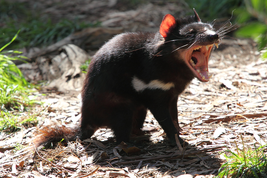
One of the mosquito species known to transmit the germ that causes malaria.

A new genetic tool may help wipe out Africa’s main malaria-carrying mosquitoes.  It’s known as a “gene drive.” In tests, it sterilized Anopheles gambiae mosquitoes. In other words, it kept them from reproducing.
A gene is the basic package of genetic instructions that tells cells how to build some protein. Proteins are the chemicals that do much of the work of cells. Gene drives are pieces of DNA that have been tweaked by scientists. These snippets of DNA have been designed to find a target gene, slice into it and then insert themselves. This cut-and-paste system alters the function of the original gene. And it is self-propagating, which means that it makes more of itself.
Other human-tweaked (or engineered) genes don’t reproduce themselves. A gene drive’s ability to make more of itself helps it get into more mosquitoes than would a regular inserted gene.
This is the second gene drive aimed at knocking out malaria. The first, announced two weeks earlier, works quite differently. It stops mosquitoes from transmitting the parasite that causes malaria. The newer gene drive instead eliminates the mosquitoes themselves by making it impossible for females to make new baby mosquitoes.
Scientists reported their achievement December 7 in the journal Nature Biotechnology.

Technique appears potent 

Most genes come in two copies. One copy comes from the mother. The other comes from the father. So each of a parent’s genes has only a 50-50 chance of getting passed on to a kid.
But like Star Trek’s Borg, gene drives become part of every unaltered target gene they encounter. These ambitious bits of DNA break standard inheritance rules. With the new technique, 76.1 percent to 99.9 percent of the offspring inherited the drive. As a result, the edited genes “drive” themselves quickly through populations.
Austin Burt first conceived of the idea for gene drives in 2003. This evolutionary geneticist works at Imperial College London, in England. For more than a decade, gene drives remained mostly just an idea. But this year, thanks to precision molecular “scissors” known as CRISPR/Cas9, four gene drives were put to use in several labs. These include the two in mosquitoes.
“They all work terrifically,” says George Church. He’s a geneticist at Harvard University in Boston, Mass.
Cas9 is a DNA-cutting enzyme borrowed from bacteria. Researchers can design genetic cousins of DNA — molecules known as RNA — to guide the enzyme to desired genes.

Not ready for prime time

Church is pleased to see that this latest mosquito gene drive works. Still, he says, further tweaks may be needed before it is ready for release into mosquitoes in the wild.
Researchers also may want to combine approaches. They might first release a gene drive that would prevent mosquitoes from carrying malaria. Later, they might release another to control mosquito reproduction, he suggests.
In the new study, Burt and colleagues first used CRISPR/Cas9 and another type of gene editor known as TALENs. These disrupted each of three genes that are very active in the egg-making organ of mosquitoes. Females carrying two copies of any one of the three disrupted genes were sterile. Once the researchers had confirmed that messing with these genes kept the mosquitoes from reproducing, the team then built gene drives to insert them into the genes.
Gene drives that interfere with reproduction make some people nervous. Such drives could make species go extinct. Many people would not miss mosquitoes. But no one really knows what getting rid of the insects might do to their ecosystem — the animals, plants and other living organisms in their neighborhood. For instance, some of those neighbors might rely on mosquitoes as lunch.
This gene drive also has some technical glitches. That means it won’t be the final version used to control wild mosquitoes. But the scientists are hopeful that future gene drives could cut populations of malaria mosquitoes dramatically, says study coauthor Tony Nolan. He’s a molecular biologist, also at Imperial College London. The public-health community needs new approaches for mosquito control, he says — “and this is a promising one.”
Cas9      An enzyme that geneticists are now using to help edit genes. It can cut through DNA, allowing it to fix broken genes, splice in new ones or disable certain genes. Cas9 is shepherded to the place it is supposed to make cuts by CRISPRs, a type of genetic guides. The Cas9 enzyme came from bacteria. When viruses invade a bacterium, this enzyme can chop up the germ's DNA, making it harmless.
cell  The smallest structural and functional unit of an organism. Typically too small to see with the naked eye, it consists of watery fluid surrounded by a membrane or wall. Animals are made of anywhere from thousands to trillions of cells, depending on their size.
bacterium (plural bacteria)   A single-celled organism. These dwell nearly everywhere on Earth, from the bottom of the sea to inside animals.
CRISPR     An abbreviation — pronounced crisper — for the term “clustered regularly interspaced short palindromic repeats.” These are pieces of RNA, an information-carrying molecule. They are copied from the genetic material of viruses that infect bacteria. When a bacterium encounters a virus that it was previously exposed to, it produces an RNA copy of the CRISPR that contains that virus’ genetic information. The RNA then guides an enzyme, called Cas9, to cut up the virus and make it harmless. Scientists are now building their own versions of CRISPR RNAs. These lab-made RNAs guide the enzyme to cut specific genes in other organisms. Scientists use them, like a genetic scissors, to edit — or alter — specific genes so that they can then study how the gene works, repair damage to broken genes, insert new genes or disable harmful ones.
disrupt     (n. disruption) To break apart something; interrupt the normal operation of something; or to throw the normal organization (or order) of something into disorder.
ecosystem   A group of interacting living organisms — including microorganisms, plants and animals — and their physical environment within a particular climate. Examples include tropical reefs, rainforests, alpine meadows and polar tundra.
enzymes   Molecules made by living things to speed up chemical reactions.
evolutionary genetics   A field of biology that focuses on how genes — and the traits they lead to — change over long periods of time (potentially over millennia or more). People who work in this field are known as evolutionary geneticists.
gene   (adj. genetic) A segment of DNA that codes, or holds instructions, for producing a protein. Offspring inherit genes from their parents. Genes influence how an organism looks and behaves.
gene editing    The deliberate introduction of changes to genes by researchers.
gene drive    A technique for introducing new bits of DNA into genes to change their function. Unlike other such genetic engineering techniques, gene drives are self-propagating. That means they make more of themselves, becoming part of every unaltered target gene they encounter. As a result, they get passed on to more than 50 percent of an altered animal’s offspring, “driving” themselves quickly into populations.
genetic  Having to do with chromosomes, DNA and the genes contained within DNA. The field of science dealing with these biological instructions is known as genetics. People who work in this field are geneticists.
genetic engineering   The direct manipulation of an organism’s genome. In this process, genes can be removed, disabled so that they no longer function, or added after being taken from other organisms. Genetic engineering can be used to create organisms that produce medicines, or crops that grow better under challenging conditions such as dry weather, hot temperatures or salty soils.
malaria   A disease caused by a parasite that invades the red blood cells. The parasite is transmitted by mosquitoes, largely in tropical and subtropical regions.
molecule (adj. molecular) An electrically neutral group of atoms that represents the smallest possible amount of a chemical compound. Molecules can be made of single types of atoms or of different types. For example, the oxygen in the air is made of two oxygen atoms (O2), but water is made of two hydrogen atoms and one oxygen atom (H2O).
organ     (in biology) Various parts of an organism that perform one or more particular functions. For instance, an ovary is an organ that makes eggs, the brain is an organ that interprets nerve signals and a plant’s roots are organs that take in nutrients and moisture.
parasite   An organism that gets benefits from another species, called a host, but doesn’t provide it any benefits. Classic examples of parasites include ticks, fleas and tapeworms.
population  (in biology) A group of individuals from the same species that lives in the same area.
proteins     Compounds made from one or more long chains of amino acids. Proteins are an essential part of all living organisms. They form the basis of living cells, muscle and tissues; they also do the work inside of cells. The hemoglobin in blood and the antibodies that attempt to fight infections are among the better-known, stand-alone proteins.Medicines frequently work by latching onto proteins.
RNA  A molecule that helps “read” the genetic information contained in DNA. A cell’s molecular machinery reads DNA to create RNA, and then reads RNA to create proteins.
sterile     An adjective that means devoid of life — or at least of germs. (in biology) An organism that is physically unable to reproduce.
TALEN   An acronym for transcription activator-like effector nucleases. These are a class of enzymes that may be used as molecular “scissors” by some gene-editing processes.
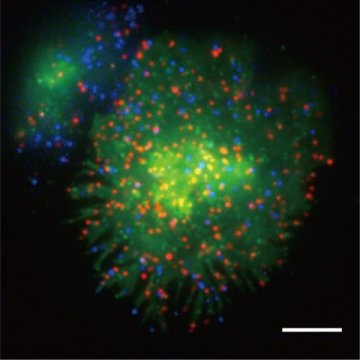